Beranda
/ Lower Body Diagram - Lower Body Diagram Fusebox And Wiring Diagram Symbol Rub Symbol Rub Sirtarghe It : The chapter on the innervation of the lower limb presents diagrams of the lumbosacral plexus and its main nerve branches for the lower limb (lateral cutaneous nerve of the thigh, femoral nerve, sciatic nerve and posterior cutaneous nerve of the thigh and obturator nerve).
Lower Body Diagram - Lower Body Diagram Fusebox And Wiring Diagram Symbol Rub Symbol Rub Sirtarghe It : The chapter on the innervation of the lower limb presents diagrams of the lumbosacral plexus and its main nerve branches for the lower limb (lateral cutaneous nerve of the thigh, femoral nerve, sciatic nerve and posterior cutaneous nerve of the thigh and obturator nerve).
Insurance Gas/Electricity Loans Mortgage Attorney Lawyer Donate Conference Call Degree Credit Treatment Software Classes Recovery Trading Rehab Hosting Transfer Cord Blood Claim compensation mesothelioma mesothelioma attorney Houston car accident lawyer moreno valley can you sue a doctor for wrong diagnosis doctorate in security top online doctoral programs in business educational leadership doctoral programs online car accident doctor atlanta car accident doctor atlanta accident attorney rancho Cucamonga truck accident attorney san Antonio ONLINE BUSINESS DEGREE PROGRAMS ACCREDITED online accredited psychology degree masters degree in human resources online public administration masters degree online bitcoin merchant account bitcoin merchant services compare car insurance auto insurance troy mi seo explanation digital marketing degree floridaseo company fitness showrooms stamfordct how to work more efficiently seowordpress tips meaning of seo what is an seo what does an seo do what seo stands for best seotips google seo advice seo steps, The secure cloud-based platform for smart service delivery. Safelink is used by legal, professional and financial services to protect sensitive information, accelerate business processes and increase productivity. Use Safelink to collaborate securely with clients, colleagues and external parties. Safelink has a menu of workspace types with advanced features for dispute resolution, running deals and customised client portal creation. All data is encrypted (at rest and in transit and you retain your own encryption keys. Our titan security framework ensures your data is secure and you even have the option to choose your own data location from Channel Islands, London (UK), Dublin (EU), Australia.
Lower Body Diagram - Lower Body Diagram Fusebox And Wiring Diagram Symbol Rub Symbol Rub Sirtarghe It : The chapter on the innervation of the lower limb presents diagrams of the lumbosacral plexus and its main nerve branches for the lower limb (lateral cutaneous nerve of the thigh, femoral nerve, sciatic nerve and posterior cutaneous nerve of the thigh and obturator nerve).. Anatomynote.com found human body artery diagram in detail from plenty of anatomical pictures on the internet. 12 photos of the muscles of the lower back and buttocks diagram. However, internally, the structure is far complex and intricate. It is usually around the size of a pea, though size varies from person to person. You need to first understand all the forces acting on the object and then represent these force by arrows in the direction of the force to be drawn.
The vertebral column of the lower back includes the five lumbar vertebrae, the sacrum, and the coccyx. The spine diagram shown below, consists of many bones or vertebrae,soft discs,the spinal cord, and spinal nerves. Between the opposing functions of the sympathetic and parasympathetic divisions of the ans, the nervous system is effectively able to control all of the organs of the abdomen and pelvis. In turn, the spinal cord relays essential information between the brain and the body. Soleus (calf muscles) tibia and fibula:
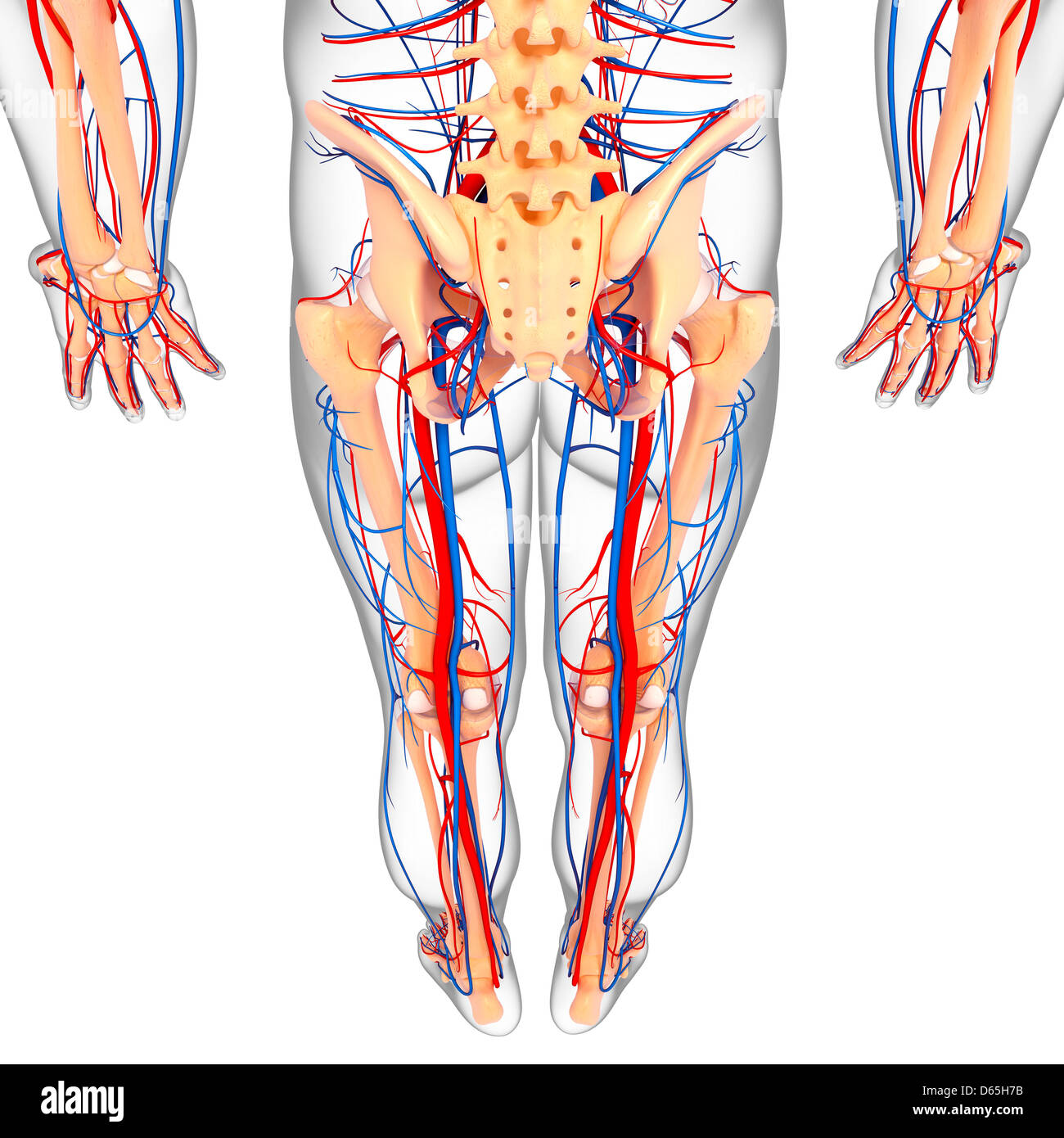
Lower Body Anatomy Artwork Stock Photo Alamy from c8.alamy.com The diaphragm forms the upper surface of the abdomen. Parasympathetic neurons in the spinal cord pass through the sacral nerves in the lower back to reach the pelvic organs such as the bladder and reproductive organs to control their functions. Proper posture and body mechanics. 12 photos of the anatomy of lower body. After receiving blood directly from the left ventricle of the heart, the. A free body diagram is defined as an illustration that depicts all the forces acting on a body, along with vectors that are applied by it on the immediate environs. The chapter on the innervation of the lower limb presents diagrams of the lumbosacral plexus and its main nerve branches for the lower limb (lateral cutaneous nerve of the thigh, femoral nerve, sciatic nerve and posterior cutaneous nerve of the thigh and obturator nerve). They provide support and a range of movements.
Anatomy of a woman's lower body, anatomy of female lower body, anatomy of lower right side of body, anatomy of the female lower body, anatomy of the lower female body, human anatomy, anatomy of a woman's lower body,.
Metatarsal # 1 (big toe) raises front of foot. Anatomy of a woman's lower body, anatomy of female lower body, anatomy of lower right side of body, anatomy of the female lower body, anatomy of the lower female body, human anatomy, anatomy of a woman's lower body,. The lumbar spine is the lower part of the back. Woman holding a blackboard with an illustration of the human digestive system drawn on it in chalk. Superficial and deep anterior muscles of upper body. The lumbar spine connects to the thoracic spine above and the hips below. Anterior and posterior muscles of the upper arm. The abdomen (commonly called the belly) is the body space between the thorax (chest) and pelvis. Between the opposing functions of the sympathetic and parasympathetic divisions of the ans, the nervous system is effectively able to control all of the organs of the abdomen and pelvis. The diaphragm forms the upper surface of the abdomen. • shift your hands down and repeat this upward motion until you reach your ankle. In turn, the spinal cord relays essential information between the brain and the body. After receiving blood directly from the left ventricle of the heart, the.
For more anatomy content please follow us and visit our website: Key bones in the abdominal area include the base of the ribcage and the lumbar spine in the lower back. The above chart notes the approximate treatment times based on the power of the device being used. The vertebral column of the lower back includes the five lumbar vertebrae, the sacrum, and the coccyx. Individual anatomical structures include 2:
Anatomy Page 55 Graph Diagram from graphdiagram.com Individual anatomical structures include 2: A book on a table • gently stretch the skin towards your upper leg and release. • shift your hands down and repeat this upward motion until you reach your ankle. Tarsals our middle foot bones. The vertebrae, which stack like spools of thread, support the back and protect the spinal cord. Labeled illustration chart on white. The diaphragm forms the upper surface of the abdomen.
A diagram shows the various inguinal lymph nodes (lymphatic ganglia).
The legs are the two lower limbs of the body. It is a diagram including all forces acting on a given object without the other object in the system. Bones, discs, and joints in your lower back. 12 photos of the muscles of the lower back and buttocks diagram. Proper posture and body mechanics. Superficial and deep posterior muscles of upper body. A book on a table For more anatomy content please follow us and visit our website: The diaphragm forms the upper surface of the abdomen. Muscles of the lower back and buttocks diagram, human muscles, muscles of the lower back and buttocks diagram. By definition, an artery is a vessel that conducts blood from the heart to the periphery. Superficial and deep anterior muscles of upper body. Each leg contains five regions.
After receiving blood directly from the left ventricle of the heart, the. These bones are connected at the back with specialized joints. Human anatomy·july 20, 2016august 22, 2016. Health conditions associated with the lower respiratory system. Ascending aorta, aortic arch, thoracic aorta, and abdominal aorta.
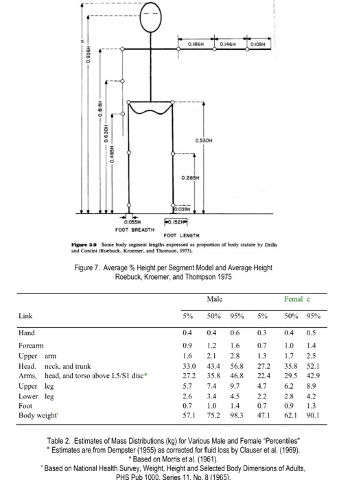
Solved Problem 2 The Free Body Diagram Of The Whole Body Chegg Com from media.cheggcdn.com Muscle diagram, most important muscles of an athletic black man, anterior and posterior view, male body. Calcaneum (by achilles tendon) raises heal when leg is bent. The spine diagram shown below, consists of many bones or vertebrae,soft discs,the spinal cord, and spinal nerves. The bones of the pelvis and lower back work together to support the body's weight, anchor the abdominal and hip muscles, and protect the delicate vital organs of the vertebral and abdominopelvic cavities. You need to first understand all the forces acting on the object and then represent these force by arrows in the direction of the force to be drawn. For your reference value these charts show the major superficial and deep muscles of the human body. We hope this picture human body artery diagram in detail can help you study and research. Soleus (calf muscles) tibia and fibula:
A free body diagram is defined as an illustration that depicts all the forces acting on a body, along with vectors that are applied by it on the immediate environs.
We think this is the most useful anatomy picture that you need. Soleus (calf muscles) tibia and fibula: Parasympathetic neurons in the spinal cord pass through the sacral nerves in the lower back to reach the pelvic organs such as the bladder and reproductive organs to control their functions. For more anatomy content please follow us and visit our website: For your reference value these charts show the major superficial and deep muscles of the human body. Stronger muscles can help stabilize the lower back and can help reduce injury risk. The diaphragm forms the upper surface of the abdomen. Proper posture and body mechanics. Pulls back scapula (shoulder blades). The lumbar spine connects to the thoracic spine above and the hips below. By definition, an artery is a vessel that conducts blood from the heart to the periphery. Veins (in blue) are the blood vessels that return blood to the heart. Anatomynote.com found human body artery diagram in detail from plenty of anatomical pictures on the internet.

.)